About Nicolas A. Karakatsanis

Nicolas Karakatsanis is appointed as Assistant Professor of Biomedical Engineering in the Department of Radiology at Weill Cornell Medical College, Cornell University, in New York city, NY. Nicolas also holds a secondary appointment as Adjunct Assistant Professor at the Translational and Molecular Imaging Institute of the Icahn School of Medicine at Mount Sinai, New York, NY. He is board certified from the American Board of Science in Nuclear Medicine with a specialty in Radiation Physics and Instrumentation. Nicolas is also a Senior IEEE member and an elected member of the IEEEE Nuclear Medical Imaging Sciences Council (NMISC). In addition, he serves as the current intern of the Physics, Instrumentation and Data Sciences Council (PIDSC) of the Society of Nuclear Medicine and Molecular Imaging (SNMMI).

A novel PET kinetics-driven bone attenuation correction method for PET/MRI

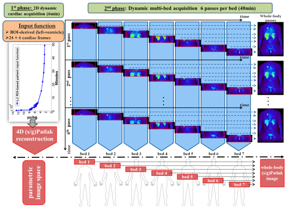
We are happy to introduce our latest method of 18F-NaF PET kinetics driven bone attenuation correction to enhance quantification of 18F-FDG and 18F-NaF images in clinical PET/MRI studies. 

In our recent article at the Journal of Nuclear Cardiology, we present a robust method for segmenting bone tissues from dynamic PET data alone, thereby allowing for the direct attenuation correction of PET data from perfectly co-registered bone attenuation maps derived from the same PET data. This capability can be important for PET/MR studies where the gold-standard method for estimating attenuation maps from transmission data is not possible.

The bone tissue maps were segmented exclusively from parametric PET images which had been derived from robust Patlak analysis of the 18F-NaF kinetics. The remaining 4 basic tissue classes of air, lungs, fat and soft tissue can be segmented from state-of-the-art MR-based tissue segmentation methods. Finally, the 5 segmented tissue classes can be combined to build a unique 5-class hybrid PET/MR-driven attenuation map to more accurately correct for PET attenuation effects.

Graph_Abstract_v2

The novel hybrid PET/MR 5-class attenuation correction method has been designed to exploit the unique kinetic properties of 18F-NaF in standalone 18F-NaF as well as dual-tracer 18F-FDG:18F-NaF PET/MR studies to enable bone tissue segmentation exclusively from PET data. The figure below demonstrates the potential benefits of the method for improving contrast and quantification of both 18F-FDG and 18F-NaF carotid images acquired with a dynamic PET/MR acquisition protocol.  

Figure9

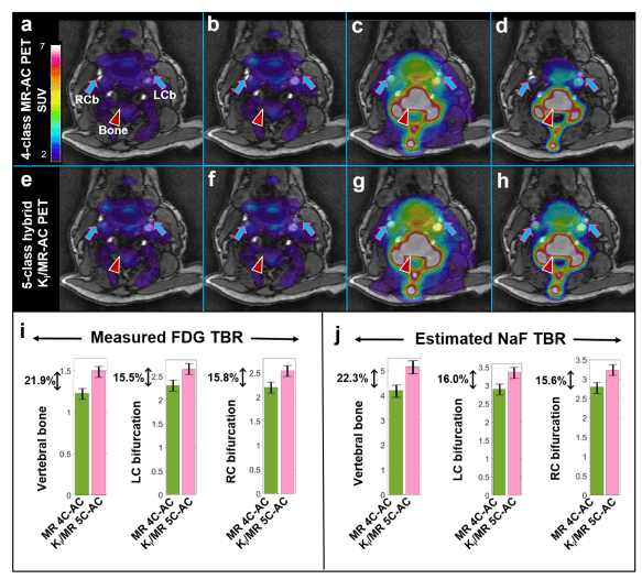
The new approach may present a robust and adoptable solution in PET/MR clinical studies to enhance PET signal quantification with two of the most widely employed PET radiotracers in cardiovascular and oncologic molecular imaging, that of 18F-FDG and 18F-NaF.


Advertisement

Pilot award on multi-parametric 68Ga-PSMA PET prostate cancer imaging

We are excited for being awarded the 2019 pilot grant from Weill Cornell Radiology Department to pursue research on whole-body (WB) multi-parametric 68Ga-PSMA PET imaging to enhance prostate cancer diagnosis and treatment response assessments.

I wish to extend my gratitude to our esteemed colleagues Dr. Scott Tagawa, Dr. John Babich, Dr. Sadek A. Nehmeh, Dr. Joseph R. Osborne and Dr. Daniel Margolis for their valuable support! I am very much looking forward to collaborating with you on this and future exciting projects.

This is an important first step towards our aim to translate dynamic WB 68Ga-PSMA PET prostate cancer imaging to the clinic.

Introducing our Nature paper on a novel nano-immunotherapy scheme for atherosclerosis

We are excited to share with you our Nature in Biomedical Engineering article on the multi-parametric evaluation of a novel nano-immunotherapy scheme targeting atherosclerosis disease. The proposed methodology moderates CD40–CD40 ligand signalling in monocytes and macrophages of atherosclerotic plaques by blocking the interaction between CD40 and tumor necrosis factor receptor-associated factor 6 (TRAF6).

The accumulation of monocytes and macrophages in atherosclerosis has been associated with destabilization and rupture of plaque which can lead to acute cardiac events. Therefore, intervening in macrophage dynamics can be an efficacious therapeutic strategy in cardiovascular diseases. Indeed, our study demonstrated that a 1-week treatment regimen achieved significant anti-inflammatory effects in mice and non-human primates, due to the impaired migration capacity of monocytes.

We are very proud to have actively participated in this wonderful journey together with more than 30 researchers in 11 departments, across three countries. If you are interested in further details, please have a look at Dr Lameijer’s news article or read our original manuscript.

Thank you!

Parametric PET imaging is now a product!

Very proud that our early work on whole-body parametric PET imaging inspired important industry vectors to translate this technology into their clinical scanner systems. We first paved the way back in 2001 at Rahmim Lab and great collaborators joined us on the way. With the support of the industry (FlowMotion MultiParametric PET by Siemens) we are now optimistic that the clinical PET community will utilize dynamic whole-body PET acquisition and direct 4D reconstruction technology to benefit from the high quantification capabilities complementing the currently established static SUV PET imaging.

Dynamic and multi-parametric whole-body PET imaging has arrived in clinic and it is readily available NOW.

Thank you!

Finally, there is a vendor product (FlowMotion Multiparametric PET by Siemens) that enables dynamic whole-body PET including parametric imaging. This is very rewarding given that our group was the earliest to propose and work on this framework, including close collaboration with Siemens. This is clearly an enabling technology, and it remains to be seen whether […]  via Rahmim Lab

Our multi-parametric PET/MR and PET/CT imaging work featured in the summer 2017 edition of the #BMEIInyc newsletter

The summer 2017 edition of the newsletter of the Biomedical Engineering and Imaging Institute (BMEII) in Mount Sinai, New York, features our  latest research findings in multi-parametric PET molecular imaging utilizing state-of-the-art:

  1. 18F-NaF kinetics-driven bone tissue segmentation,
  2. dual-tracer PET/MR carotid plaque imaging and
  3. combined SUV/Patlak-4D WB PET/CT oncologic imaging

Download the newsletter in PDF format here.

TMIInewsletter_Summer2017_Page3

Page 3 of the TMII newsletter featuring our work in multi-parametric PET/MR and PET/CT

Our whole-body 4D parametric PET imaging research featured in 2016 highlights of the PMB journal

We are grateful to announce that our recent research publication in direct 4D whole-body PET parametric imaging has been featured in 2016 highlights of the Physics in Medicine and Biology (PMB) journal. Please check below the line-up of highlighted 2016 PMB articles awarded by the editorial team of the prestigious PMB journal in the field of PET imaging:

http://iopscience.iop.org/journal/0031-9155/page/Highlights_of_2016

This is indeed a great honor and we are indebted to the great interest you have recently demonstrated for our on-going progress in this new exciting molecular imaging field.

Our work aims at translating the quantitative virtues of multi-parametric molecular PET imaging, as initially had been shown in neuroimaging, to clinically feasible multi-bed PET scan protocols. The transition to whole-body dynamic and parametric PET imaging is critical as it may substantially enhance the diagnostic, prognostic and theranostic prospects of image assessments of various important molecular mechanisms  of oncologic and cardiovascular diseases.

figure1_gpatlak4dpaper

Direct 4D whole-body dynamic PET parametric imaging method

Thank you all for your active support and we would like to take this opportunity to openly share once again  our continuous commitment to advance the clinical value of molecular PET imaging as a valuable biomedical imaging tool to assess progress and therapy responses at the molecular level.

Our paper on direct 4D Whole-Body PET parametric imaging in the Top 10 PMB articles for 2016!

We are grateful to announce that our featured article on direct-4D whole-body PET parametric image reconstruction has been included in the Top-10 rankings for the most popular Physics in Medicine and Biology (PMB) articles of the year!

This is indeed a honorary distinction, as it stems directly from the broad interest of nuclear medicine community in our vision for clinically adoptable dynamic and parametric PET imaging.

Therefore, we would like to take this opportunity and thank you for your continuous feedback and support in our on-going efforts to enhance the quantitative virtues of PET molecular imaging by translating dynamic and parametric 4D PET imaging to the clinic.

In case you have missed the article, you can find it via the following top-10 PMB articles list for 2016, among other exciting scientific work in related fields.

Top-10 Most Popular Physics in Medicine and Biology Articles for 2016  (Offline View)

Article on the clinical potential of whole body dynamic 4D PET imaging

We are very excited and motivated that our recently published work in dynamic whole-body 4D PET imaging for the clinical translation of advanced quantitative PET imaging has began to take notice in scientific community, as seen in the following online news article:

A clinical take on whole-body dynamic PET   (Offline Access)

The presented developments from Dr Karakatsanis and colleagues are the fruits of the productive collaboration between the research groups of Dr Rahmim at Johns Hopkins University and Dr Zaidi at University of Geneva.

The research work is further advanced currently at Icahn School of Medicine at Mount Sinai, New York, in close collaboration with the previous two groups.